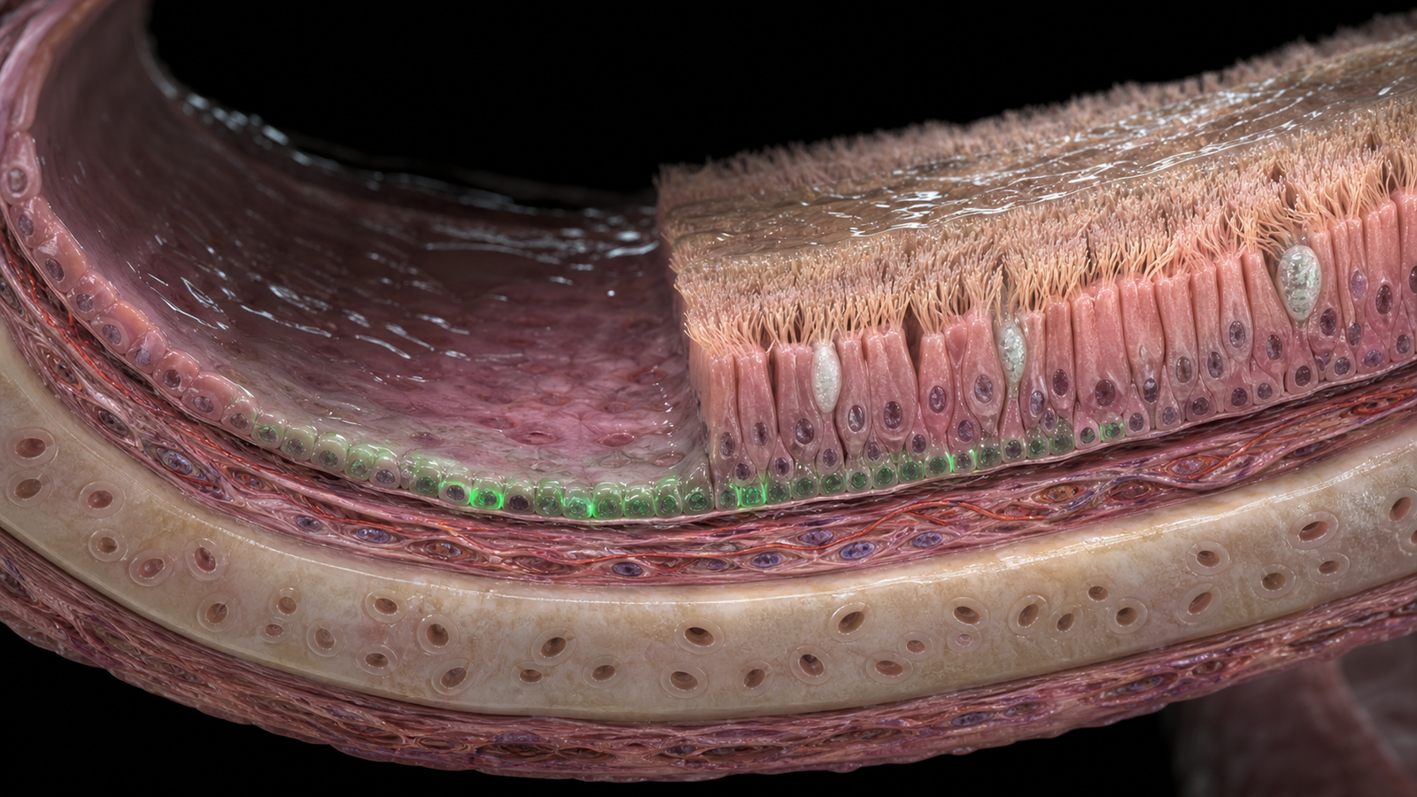
Editorial illustration of donor basal cells integrating into an injured airway and rebuilding a ciliated epithelial lining. Credit: CellXperience generated editorial illustration.
Under the microscope, a successful airway graft should eventually stop looking like a graft. Donor basal cells have to settle into the basal layer, then leave behind ciliated and secretory descendants that move chloride and mucus. A new JCI Insight research letter reaches that point in ferret experimental systems, one step before a living-animal test.
The experiments are small. Their functional measurements make the paper unusually informative.
Primary sources
- JCI Insight, "Basal cell transplantation supports multilineage differentiation and restores CFTR function in ex vivo ferret airways" - https://insight.jci.org/articles/view/206375
- Cell Stem Cell, "Airway stem cell reconstitution by the transplantation of primary or pluripotent stem cell-derived basal cells" - https://pmc.ncbi.nlm.nih.gov/articles/PMC10528754/
The airway had to be injured before donor cells could enter
Airway epithelium is built to exclude foreign material. Ciliated and secretory cells cover the basal stem-cell compartment, mucus traps incoming particles, and innate defenses remove them. Those protections become obstacles when the incoming material is a therapeutic cell.
The researchers first had to create an open niche. They grew primary ferret bronchial epithelial cells at an air-liquid interface, producing a polarized lining with its apical side exposed to air. Wild-type and cystic-fibrosis G551D cultures were treated with polidocanol to damage part of the epithelium.
Too much polidocanol killed the culture. Too little preserved the barrier so well that donor cells had nowhere to implant. At 0.1 percent, about 59 percent of cells remained alive while transepithelial electrical resistance fell by 95 percent. The damaged cultures later restored their barrier, making that concentration useful for transplantation experiments.
Conditioning is the method's first hard problem. A therapeutic airway cannot simply be flooded with replacement cells and expected to accept them. Space has to be created without causing an injury worse than the disease being treated.
Wild-type cells occupied most of the CF culture
The team labeled wild-type ferret bronchial epithelial cells with GFP and placed 150,000 of them onto injured G551D cystic-fibrosis cultures. Four weeks later, donor-derived cells made up about 77 percent of the epithelium.
The rebuilt layer was tested in two ways that connect directly to cystic-fibrosis physiology. Ussing-chamber measurements showed recovery of CFTR-dependent chloride current. Micro-optical coherence tomography showed mucociliary transport at about 1.2 millimeters per minute, close to the 1.1 millimeters per minute measured when wild-type cells were transplanted onto wild-type controls. Injured CF cultures without donor cells remained functionally poor.
These are persuasive measurements inside a controlled culture system. CFTR function was restored after healthy cells took over much of a CF epithelial layer, and the surface moved mucus again. Each condition contained three replicates, so the result needs replication at a larger scale before anyone can treat the percentages as stable performance estimates.
The donor cells were primary wild-type ferret cells. They were neither human cells nor gene-corrected cells from an affected recipient. A future autologous therapy would have to collect or derive basal cells, correct the disease-causing variant, expand them without losing stem-cell behavior, and deliver enough viable cells back to the airway.
The excised trachea answered a different question
The researchers then used whole pieces of wild-type ferret trachea maintained outside the animal. They brushed the luminal surface about 15 times to remove part of the existing lining, seeded 20 million GFP-labeled wild-type cells, and cultured the tissue for three weeks.
Donor cells made up roughly 30 percent of the surface epithelium. They had produced ciliated cells, MUC5B-positive secretory cells, and KRT5-positive basal cells. The grafted tissue showed CFTR-dependent ion transport and active cilia.
Functional rescue of the G551D defect belongs to the air-liquid-interface cultures. The excised tracheae were healthy wild-type tissue and tested whether cells could engraft, diversify, and contribute to a functioning lining in intact airway architecture. No living ferret received the transplant in this paper.
Disease-function correction came from simplified cultures, while intact tracheal tissue supplied the evidence for multilineage reconstruction.
Basal cells carry the right job description
Basal cells are resident stem cells of the conducting airway. They self-renew and produce ciliated and secretory descendants during normal maintenance and repair. A successful transplant could therefore install a renewable source of corrected epithelium instead of supplying mature cells that disappear after a short period.
Earlier work from overlapping investigators showed that primary or pluripotent-stem-cell-derived basal cells could reconstitute injured mouse airways. Mouse donor cells persisted for more than two years and retained self-renewal through serial transplantation. Human primary and pluripotent-derived basal cells also formed multiple airway lineages after transplantation into immunodeficient mice.
The ferret work adds a disease-relevant functional test. Ferret airway physiology and cystic-fibrosis phenotypes are closer to the human condition than many mouse readouts, and the investigators measured chloride transport and mucus movement rather than relying on fluorescent engraftment alone.
Engraftment still depends on controlled injury
Both experiments opened the niche deliberately. Polidocanol damaged the cultured epithelium. Mechanical brushing removed cells from the tracheal surface. Neither procedure is ready for diffuse treatment of a person's airways.
Airway geometry turns this into a delivery problem. A clinical protocol would have to reach a large, branching surface coated in mucus, avoid excessive inflammation, keep the airway open, and expose enough basal territory for donor cells to attach. Existing disease may create damaged patches, but it cannot be assumed to provide uniform or controllable access.
Immune compatibility adds another decision. Autologous gene-corrected cells may reduce rejection while demanding individualized manufacturing and extensive genomic quality control. A banked allogeneic product would simplify supply and bring immune recognition back into the procedure. Pluripotent-derived basal cells could expand the starting material, with residual pluripotency and differentiation consistency requiring their own safety assays.
The functional benchmark is now visible: enough donor-derived epithelium to restore CFTR current and move mucus in a ferret system. Delivery, conditioning, and cell source remain open engineering choices.
A living ferret will be much less forgiving
A living ferret adds blood flow, immune surveillance, breathing forces, infection, mucus accumulation, coughing, and continuous epithelial turnover. Donor cells will have to survive delivery through that environment and remain useful after the conditioning injury has healed.
The next experiment should determine how much of the airway must be replaced for a durable physiological benefit, whether corrected cells retain their advantage over time, and whether conditioning can be localized without creating dangerous denudation. Cell distribution across the branching airway will matter as much as the average engraftment percentage in a sampled region.
The decisive result will be a living airway that keeps donor-derived epithelium, transports mucus, and resists the return of cystic-fibrosis dysfunction after recovery from the transplantation procedure.