
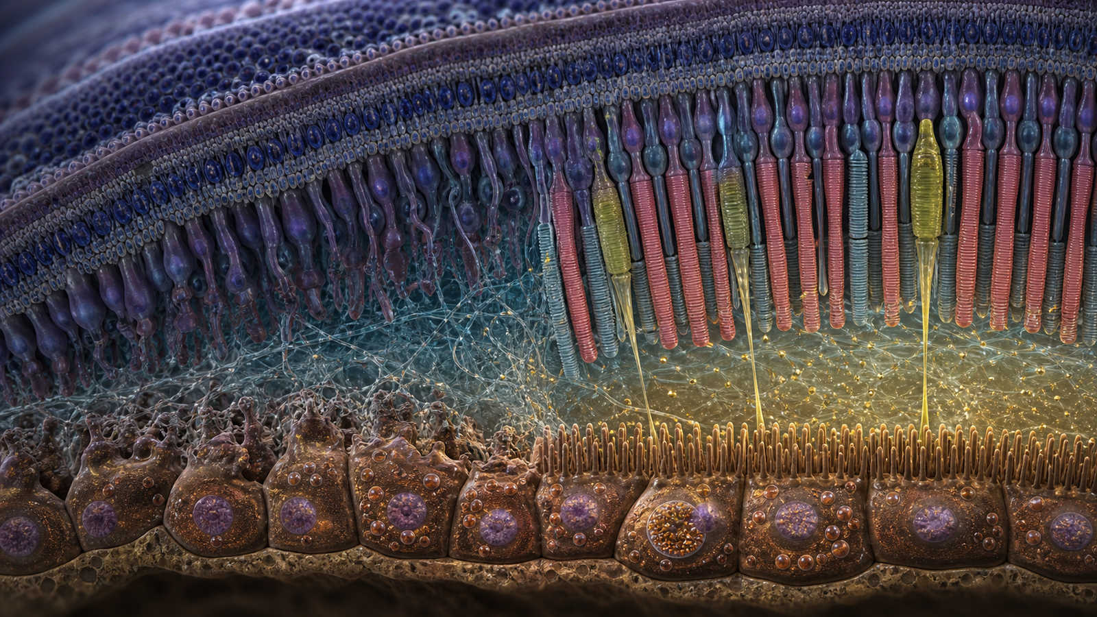
Editorial illustration of the retinal organoid-RPE interface used to study CLN3 disease and acid ceramidase rescue. Credit: CellXperience generated editorial illustration.
Photoreceptors cannot maintain their light-sensing outer segments alone. They shed part of this material every day, and the retinal pigment epithelium beneath them clears the debris while supporting renewal. CLN3 disease disrupts that partnership early, but conventional retinal organoids leave out the mature epithelial interface needed to ask which side fails first.
A new Science Translational Medicine study builds the missing contact. Human stem-cell-derived retinal organoids sit in a degradable matrix above a pigmented RPE layer, allowing their outer segments to reach the supporting cells. When the researchers exchanged healthy and CLN3-mutant components, mutant RPE damaged control photoreceptors. Mutant photoreceptors remained comparatively organized when the RPE was healthy.
The experiment shifts the origin of early retinal damage away from an exclusively neuronal explanation. It also identifies acid ceramidase as a possible point of intervention, then tests recombinant enzyme in the human model and in a CLN3 miniswine.
Primary sources
- Science Translational Medicine, "A 3D human retina model reveals a non-cell-autonomous and non-neuronal mechanism of photoreceptor loss in a lysosomal storage disorder" - https://www.science.org/doi/10.1126/scitranslmed.ady7616
- Complete exact-title bioRxiv manuscript - https://www.biorxiv.org/content/10.1101/2025.07.10.664233v1.full
Science Translational Medicine published the peer-reviewed report on August 26, but its full text was not openly accessible when this article was prepared. Procedural and numerical details below therefore come from the authors' complete bioRxiv manuscript; the journal abstract reports the same central model, mechanism, animal work, and rescue experiments.
Rebuilding the photoreceptor-RPE contact
Retinal organoids can produce layered neural retina with rods and cones, but their photoreceptor outer segments often face culture medium rather than a functional RPE surface. RPE monocultures provide the opposite half, modeling phagocytosis and lysosomal biology without recreating the physical relationship to photoreceptors.
The investigators joined the two. Human pluripotent stem-cell-derived retinal organoids were placed in a polyethylene glycol and hyaluronan matrix. A peptide that matrix metalloproteinases can degrade crosslinked the material. The construct sat above an hPSC-derived RPE monolayer in a transwell arrangement.
Rather than serving only as a scaffold, the matrix creates a permissive space resembling the interphotoreceptor matrix, through which outer segments can extend toward RPE microvilli. The RPE can then contact, engulf, and process shed photoreceptor material.
After seven days of co-culture, control organoids paired with control RPE maintained organized rod and cone outer segments. This provided a short experimental window for changing either the neural or epithelial genotype while holding the other side constant.
The decisive pairing uses healthy photoreceptors
CLN3 disease, also called juvenile neuronal ceroid lipofuscinosis or juvenile Batten disease, is a lysosomal storage disorder. Children develop progressive vision loss. Neurological decline follows. The common deletion of exons 7 and 8 in CLN3 affects many cell types, making it difficult to determine whether photoreceptors degenerate because they carry the mutation or because their support system fails.
The researchers derived retinal organoids and RPE carrying that deletion. A culture made from mutant organoid and mutant RPE showed disorganized and depleted photoreceptor outer segments, reproducing an important feature of the disease.
The mixed cultures localized the problem. Healthy retinal organoids developed outer-segment loss when paired with mutant RPE. Mutant organoids cultured without mutant RPE retained outer segments for extended periods, including observations reported up to a year. A genetic defect within the RPE was therefore sufficient to injure photoreceptors that did not carry it.
Mutant RPE cells develop their own lysosomal and structural abnormalities, making the CLN3 defect cell autonomous within that tissue. Photoreceptor injury is non-cell-autonomous because it arises in a neighboring cell population through failure of epithelial support.
A photoreceptor-only treatment may miss the earliest actionable compartment. Restoring RPE handling of outer segments could preserve neurons even if the inherited CLN3 mutation remains present throughout the retina.
Acid ceramidase connects lysosomes to the epithelial surface
The mutant RPE showed reduced acid ceramidase, an enzyme that breaks down ceramide inside lysosomes. Sphingosine produced through that pathway can be converted to sphingosine-1-phosphate, a signaling lipid involved in membrane dynamics and cell structure. Both acid ceramidase and sphingosine-1-phosphate were reduced in the CLN3 system.
The consequences appeared at the apical surface of the RPE. EZR, also known as ezrin, normally helps link the cell membrane to the actin cytoskeleton in RPE microvilli. Those microvilli wrap around photoreceptor outer segments and support their uptake. In mutant RPE, EZR localization and microvillar organization were disturbed.
Outer-segment phagocytosis also declined. The RPE was present and pigmented, yet its lysosomal lipid metabolism and the surface machinery that meets photoreceptors no longer worked together normally. The paper therefore connects a lysosomal storage mutation to a physical failure at the tissue boundary.
This route is plausible without being exclusive. CLN3 affects trafficking, autophagy, metabolism, and other lysosomal functions. Acid ceramidase may sit within a larger network of abnormalities rather than serving as the single driver of retinal disease. The compartment-swapping result establishes the importance of RPE more firmly than it establishes one complete molecular chain.
Human eyes and miniswine support the model
The final paper reports examination of donor eyes from two people with CLN3 disease. Structural and molecular changes in those tissues agreed with observations from the stem-cell model. High-resolution retinal imaging in two living patients also suggested reduced RPE autofluorescence early in disease.
Four human cases across donor tissue and living imaging cannot define how consistently the pattern appears, when it begins, or how it relates to later visual decline. They serve as rare corroboration in a disease where access to early retinal tissue is extremely limited.
CLN3-mutant miniswine provided a larger eye with retinal anatomy closer to humans than a mouse eye. The investigators observed disorganized photoreceptor outer segments and altered RPE microvilli in the central retina of young animals. Those findings extend the organoid-RPE phenotype into a living system with normal ocular architecture and circulation.
The miniswine model still differs from children with CLN3 disease, and the reported abnormalities emerged within an accelerated experimental timeline. Its value comes from testing delivery and tissue response in an intact eye, not from reproducing every stage of human disease.
Recombinant enzyme improves structure over seven days
The authors treated the organoid-RPE cultures with recombinant human acid ceramidase, or rhAC. In the complete manuscript, cultures received 30 micrograms per milliliter for one hour daily. Treatment continued for seven days. Acid ceramidase activity rose, and rod and cone outer-segment organization improved.
They then moved rhAC into the miniswine eye, giving intravitreal enzyme to one eye and vehicle to the other. After seven days, treated eyes showed higher acid ceramidase, lower ceramide, improved EZR localization, and better-preserved photoreceptor outer-segment structure.
Only three biological replicates were used for this intervention. Some findings were qualitative. The reported ceramide comparison approached rather than crossed the conventional statistical threshold. The study did not show improved vision, durable rescue, repeated-dose tolerability, or preservation across months.
A short mouse experiment found no obvious fundus, angiography, or electroretinography abnormality seven days after ocular dosing. That check addresses immediate ocular toxicity at the tested time point, not a chronic safety profile for an enzyme that may require repeated administration.
Replacing the deficient enzyme changed the proposed pathway and improved the tissue structure expected to depend on it. The experiment tests causality more directly than a correlation can, while remaining far earlier than a demonstration of therapy.
From an eye experiment to a treatment program
Intravitreal enzyme delivery has an established clinical precedent in ophthalmology, which makes the route imaginable. CLN3 disease adds difficult questions. The enzyme must reach the relevant RPE compartments at an active concentration, persist long enough to matter, and avoid inflammation or toxicity after repeated dosing. A treatment for the eye would also leave the neurological progression of CLN3 disease unresolved.
The human stem-cell model can help answer dose and mechanism questions before larger animal studies. Because healthy and mutant components can be exchanged, researchers can test whether treatment must reach the RPE, photoreceptors, or both. The same arrangement can reveal whether outer-segment rescue continues after enzyme withdrawal and whether intervention still works once disorganization is established.
The next translational threshold is a blinded, adequately powered miniswine study that follows retinal structure and visual function for months after repeated dosing. Without that durability, seven-day structural improvement remains an elegant experiment rather than a therapeutic result.