
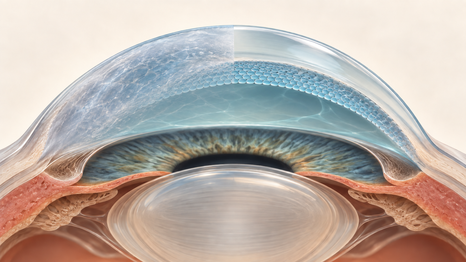
Generated editorial illustration of cultured endothelial cells forming a layer on the posterior corneal surface. Credit: CellXperience.
The corneal endothelium is a single layer of cells with an outsized job. It pumps fluid out of the corneal stroma, helping the tissue remain thin and transparent. When that layer fails, the cornea swells and vision clouds. Conventional treatment replaces donor tissue. Cultured corneal endothelial cell therapy instead delivers a cell suspension into the eye and asks those cells to rebuild the inner surface.
A report published in Cornea on September 21 follows two people who had already taken part in the first human study of this approach. Their prototype cell grafts initially cleared the cornea, then lost function four and nine years later. Both eyes received a newer mature-cell product in 2024. One year after the second treatment, each eye again had a measurable endothelial layer and a thinner cornea.
The report is small enough to read as two clinical histories rather than an efficacy study. Its value lies in a narrower question: can a cornea that has already received cultured endothelial cells be prepared and treated again?
A suspension replaces a tissue graft
The treatment, marketed in Japan as Vyznova, combines one million cultured allogeneic corneal endothelial cells with the Rho kinase inhibitor Y-27632. Before injection, the surgical team polished an 8-millimeter central zone of the recipient's Descemet membrane to remove degenerated cells and abnormal extracellular matrix. The cell suspension was then injected into the anterior chamber.
Patients remained face down for three hours so that gravity would help the cells contact the posterior cornea. Systemic corticosteroids were used for one week, followed by an extended course of topical corticosteroid and antimicrobial drops. The procedure therefore involves a cell product, surgical preparation, positioning, and postoperative immunosuppression. The injection cannot be evaluated apart from that complete workflow.
Once attached, endothelial cells form the pump layer that regulates corneal hydration. That makes central corneal thickness, endothelial cell density, and visual acuity relevant follow-up measures. It also makes loss of pressure control, inflammation, and rejection important safety observations.
The first products worked, then declined
Both participants received prototype cultured cells in 2014. One had pseudoexfoliation syndrome. That eye remained clear for about four years, then developed endothelial failure and persistent edema for the next six. The other participant had Fuchs endothelial corneal dystrophy. Her graft retained useful function longer, with cell density falling to roughly 500 cells per square millimeter from year four onward and endothelial failure occurring around year nine.
The authors connect those late failures to the maturity profile of the early cell preparations. Their manufacturing program uses a surface-marker combination to identify mature differentiated endothelial cells and summarizes the proportion with an “E-ratio.” The 2014 prototype lots had a lower E-ratio than the later product.
This is a biologically plausible product-quality explanation supported by the group's earlier development work. The two retreatment cases cannot prove why the original grafts failed. They also cannot isolate maturity from disease, postoperative conditions, or other patient-specific influences across a decade.
One year after retreatment
The first eye entered retreatment with a central corneal thickness of 786 micrometers. At one year it measured 572 micrometers, with an endothelial cell density of 2,292 cells per square millimeter. Decimal best-corrected visual acuity improved from 0.03 to 0.4. The authors still observed mild stromal haze, which they relate to the six years of persistent edema before retreatment.
The second eye changed from 688 to 523 micrometers in central thickness and reached 2,552 endothelial cells per square millimeter at one year. Decimal visual acuity improved from 0.07 to 1.0. That eye had spent much less time in a failed, edematous state before the second procedure.
Neither participant developed graft rejection, elevated intraocular pressure, or anterior uveitis during the reported year. Those observations are useful for these two eyes. A sample of two cannot estimate uncommon adverse events, compare retreatment with endothelial keratoplasty, or establish a general safety rate.
Timing may matter after the first graft fails
The contrast between the cases gives the paper its most practical signal. Cell replacement restored the pump layer in both eyes, while the eye exposed to six years of edema retained more stromal haze and less visual improvement. Endothelial recovery can remove excess fluid. It may not reverse every structural consequence of prolonged swelling.
That interpretation remains observational. The participants had different underlying diseases, and the study did not assign retreatment timing. Still, it frames a useful follow-up question for future cohorts: once endothelial failure is confirmed, does earlier retreatment preserve a clearer optical path?
Durability is the other unresolved issue. The first grafts had looked successful at one year too. The mature product differs from those prototypes, and earlier work from the same program associates its maturity profile with better outcomes. Only longer observation will show how its cell density changes after a second injection.
What this report changes
Repeat treatment is no longer only a theoretical extension of cultured endothelial cell therapy. The team cleared the recipient surface, delivered a mature-cell product, and observed a functioning endothelial layer one year later in two previously treated eyes. That is a technical and clinical feasibility result.
Broader conclusions need a larger, deliberately followed retreatment cohort. Useful next data would include prespecified failure criteria, time from failure to retreatment, product release characteristics, longer cell-density curves, and comparison with repeat tissue keratoplasty. Etiologies beyond pseudoexfoliation syndrome and Fuchs dystrophy also need representation.
The scale of the report should stay visible beside the attractiveness of the method. Two successful one-year retreatments show that the inner corneal surface can accept another cultured-cell layer. They do not yet tell clinicians how often it will work, how long the second layer will last, or which patients should receive it instead of donor tissue.
Source
The analysis uses the complete open article, “Safety and Efficacy of Mature Cultured Corneal Endothelial Cell Therapy (Vyznova) in Eyes With Declining Long-term Efficacy After Prototype Cell Transplantation”, published online September 21, 2026. The report describes two observational retreatment cases with one year of follow-up. The hero is an original generated editorial illustration and does not depict either participant or measured study data.