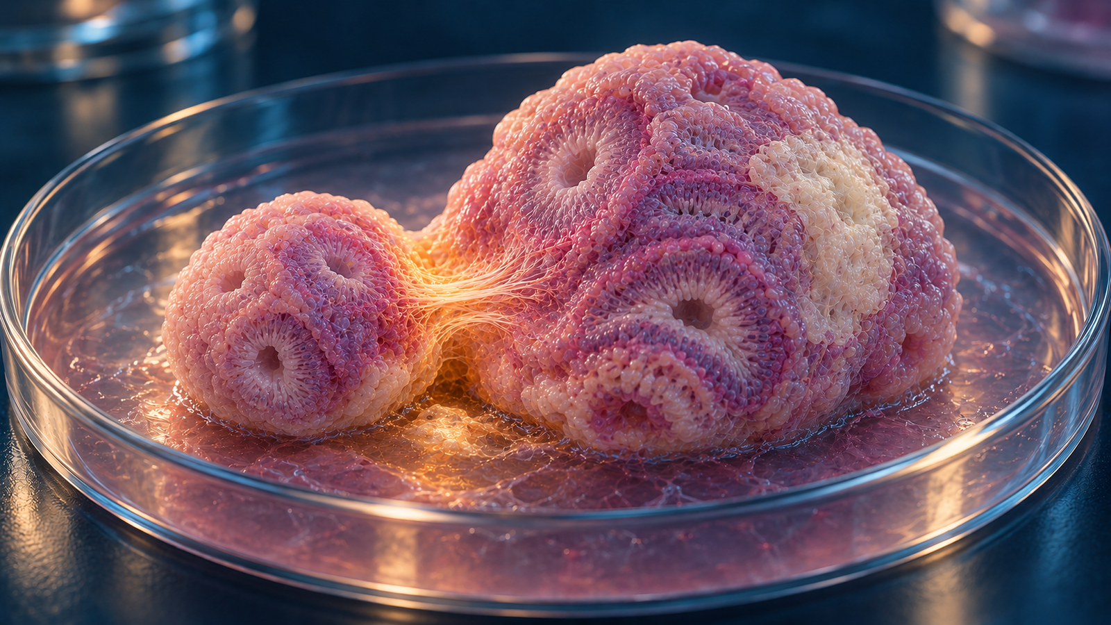
Generated editorial illustration of an early cortical organoid attaching to injured mature organoid tissue in culture. Credit: CellXperience.
Most transplantation studies place dissociated cells or a tissue fragment into an animal. A bioRxiv preprint posted October 1 uses a more controlled pairing: one human cortical organoid serves as the graft and another as the injured host. The model lets researchers watch two pieces of neural tissue meet, adhere, and exchange cells and fibers without the many variables introduced by surgery and a living brain.
The donor organoid was developmentally young and rich in proliferating neural progenitors. The host was older, more neuronal, and exposed to six hours of oxygen-glucose deprivation. A small molecule called ER272 was added for seven days to expand the donor’s progenitor pool and alter programs involved in adhesion and extracellular matrix. By two weeks, the combination produced more integration and neuronal repair markers than transplantation or drug treatment alone.
It is an elegant reduction of the graft-host interface. It is also a preprint built from immature tissue in a dish. Its relevance depends on keeping both facts visible.
Two organoids take different roles
The researchers generated cortical organoids from human pluripotent stem cells. Early organoids were labeled with green fluorescent protein so that donor-derived cells and fibers could be separated from the unlabeled host. Mature organoids were allowed to develop longer before injury.
Age mattered. About 20% of cells in the early organoids expressed the proliferation marker Ki67, compared with roughly 5% in the later organoids. The younger tissue supplied a reservoir that could still expand and generate neurons. The older tissue better represented a differentiated neural target, though it remained far from an adult cortex.
The host organoid underwent oxygen-glucose deprivation, a laboratory stress used to reproduce part of ischemic injury. Six hours of deprivation caused neuronal loss and reduced markers of cortical identity and maturation. The team then placed one early donor organoid against the injured host and maintained direct contact between them.
This “organoid-to-organoid” transplant preserves tissue architecture on both sides of the interface. Donor cells arrive with neighbors, extracellular matrix, and radial organization instead of as a suspension. The host offers a three-dimensional target with injured neurons and local matrix. That makes the system useful for asking how tissues fuse, while leaving out blood flow, immune surveillance, and long-range connectivity.
ER272 acts on the younger tissue
ER272 was developed as a pro-neurogenic small molecule. In this study, it activated protein kinase C alpha and beta-1 and increased Notch signaling. The early organoids responded with a larger pool of proliferating neural progenitors. Late organoids had fewer responsive progenitors, which helps explain why ER272 alone did not rescue the injured host.
That distinction sharpened the experimental logic. A drug can fail against mature injured tissue and still be useful when applied to a graft that contains the cell state it can influence. Here, ER272 appears to prepare the donor organoid for expansion and integration rather than directly reversing damage in the host.
Transcriptomic analysis supported that interpretation. Treated donor tissue increased genes associated with extracellular matrix organization, cell adhesion, integrins, and growth-factor signaling. Those changes could help cells remain at the interface, enter the host, and extend processes through it. They are associations from gene-expression data, not proof that any one pathway is necessary for the observed integration.
Integration rises within a week
Seven days after transplantation, about 90% of the ER272-treated donor organoids had integrated with their hosts. The untreated control and oxygen-glucose-deprivation groups were near 40%. Integration was judged from the physical union of the tissues and movement of fluorescent donor material into the host.
The drug also expanded the graft. Treated early organoids contained more Ki67-positive green cells and a larger donor-derived area. In the authors’ summary of the developmental comparison, early organoids contained about twice as many total cells and 3.5 times as many newly generated CTIP2-positive deep-layer neurons as late organoids.
These measurements show that the treatment changed the donor compartment. They do not yet separate beneficial integration from simple overgrowth. A transplant intended for the brain would need a reliable endpoint to proliferation, predictable final composition, and a sensitive search for residual pluripotent or aberrant cells.
Neurons and fibers enter the injured host
By day 14, treated grafts had produced more donor-derived neurons and fibers inside the host organoid. The authors measured increases in HuC/D, a neuronal marker; CTIP2, associated with deep-layer cortical neurons; and MAP2, found in neuronal dendrites. Across the combined model, several neuronal signals returned toward levels in uninjured controls.
The green label made the origin of invading cells and processes visible. Donor fibers traversed the boundary and extended into the older tissue. Host-level marker recovery suggests that the paired system contained more neuronal material after treatment, but it does not show that those fibers formed functional synapses or restored a network computation.
Electrophysiology would be an important next step. Spontaneous activity, evoked responses across the graft-host boundary, and selective interruption of donor activity could reveal whether the new processes participate in a circuit. Longer culture would show whether the initial bridge matures, stabilizes, or becomes disorganized.
A useful model of one difficult boundary
Neural graft studies struggle to attribute failure. Cells may die during injection, encounter a hostile extracellular matrix, lack trophic support, face immune attack, or fail to connect with the right targets. An organoid pair cannot reproduce that full landscape. It can make one part of it experimentally accessible.
The model is especially suited to the physical and molecular boundary between donor and host. Researchers can vary donor age, host injury, contact geometry, matrix composition, and treatment timing. They can image the same preparation repeatedly and recover each compartment for molecular analysis.
The omissions are substantial. Organoids lack a perfused vascular system and the blood-brain barrier. They contain limited immune and glial diversity, no meninges, and no normal input from other brain regions. Oxygen-glucose deprivation creates an abrupt metabolic insult without the vascular, inflammatory, and mechanical features of a human stroke. Fourteen days cannot address long-term graft maturation, seizure risk, tumorigenicity, or behavior.
The authors’ use of “repair” is supported at the level of tissue integration, neuronal markers, and fiber growth. It should not be stretched to clinical recovery. The experiment does not establish a transplant dose, surgical route, immunological strategy, or safety profile.
Where the method can go next
The most direct follow-up is mechanistic. Blocking selected integrins, matrix components, or Notch activity could show which ER272-induced changes are required for integration. Live imaging could distinguish migration, local cell division, and fiber extension. Single-cell measurements across time could reveal whether treatment alters neuronal subtype balance or leaves an unusually proliferative population behind.
A more complete model could add microglia, vascular cells, or an immune-compatible barrier. The donor could also be compared with dissociated neural progenitors, engineered tissue strips, or organoids of different ages. Such experiments would clarify whether preserved tissue architecture provides an advantage beyond delivering more progenitors.
Animal transplantation would remain a separate test. A graft that fuses with another organoid may not survive injection, orient within a lesion, connect over long distances, or tolerate an immune response. The current system is valuable precisely because it does not pretend to answer all of those questions at once.
The preprint shows a younger neural tissue responding to a drug, joining an injured older tissue, and sending neurons and fibers across their boundary. That is a credible platform result. The path from that interface to a repaired human cortex is still long and experimentally undefined.
Source
This analysis uses the complete version 1 bioRxiv preprint, “Organoid-to-organoid transplantation and pro-neurogenic treatment promote graft integration and neuronal repair in a human ischemic model”, posted October 1, 2026. The manuscript had not undergone peer review at the time of publication and declares no competing interests. The hero is an original generated editorial illustration, not microscopy or a figure from the study.