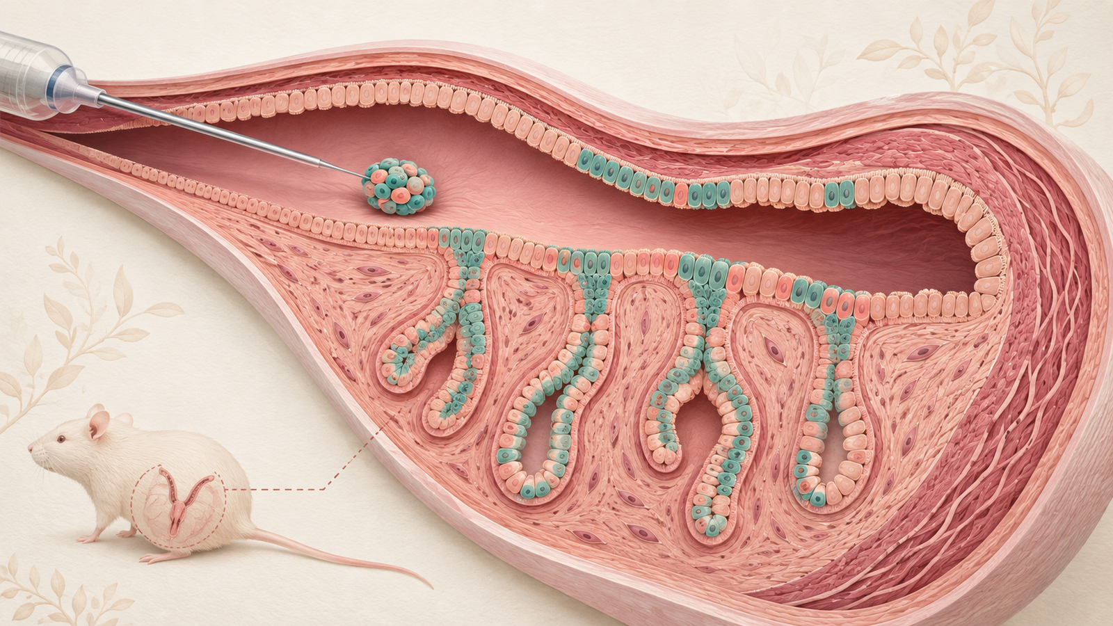
Editorial illustration of endometrial organoid cells rebuilding luminal and glandular uterine epithelium. Credit: CellXperience generated editorial illustration.
The cleanest result in this study is anatomical. One uterine horn received endometrial organoid cells. The other received vehicle. After the injured mice recovered and mated, implantation occurred in the transplanted horns and not in their untreated partners. The pregnancies continued to live birth.
That within-animal comparison turns fertility into a demanding graft readout. Donor cells had to survive delivery, occupy the correct epithelial compartments, respond to ovarian and embryonic signals, and support implantation long enough for pregnancy to progress. A lining that merely looked repaired would not have passed the test.
The experiment also revealed something less expected about cell identity. When the investigators removed glandular-lineage cells from donor organoids before transplantation, the remaining luminal-lineage cells formed new glands inside the uterus. The adult tissue environment induced a fate that extended culture did not.
Primary source
The complete manuscript was posted August 31, 2026, under a CC BY 4.0 license. It is a preprint and has not been certified by peer review. All procedural, numerical, limitation, and figure-level details in this analysis come from that version.
Re-covering the uterine surface did not restore function
The endometrium contains a luminal epithelium, the surface facing the uterine cavity, and glandular epithelium that extends into the underlying stroma. Both compartments change across reproductive cycles. The glands secrete factors needed for embryo implantation and early pregnancy, so reconstructing a continuous surface cannot substitute for rebuilding the whole epithelial system.
The researchers created a genetic injury to expose that distinction. Female mice expressed a diphtheria-toxin receptor in uterine epithelial cells under control of an Ltf-iCre system. One toxin dose triggered epithelial apoptosis while preserving the underlying stroma. Flow cytometry and histology estimated that about 94% of the epithelial population was lost.
By day ten, cells that escaped ablation had spread across the denuded stroma and re-covered the luminal surface. The repair looked substantial at low resolution. Yet the mice failed to establish pregnancies during a three-month breeding assessment. The regenerated surface had not reconstructed enough glandular architecture or function to support fertility.
This model is deliberately severe and precise. It removes epithelial cells through engineered toxin sensitivity rather than inflammation, fibrosis, surgery, infection, or postpartum injury. That precision helps isolate the epithelial contribution. It also makes the injury unlike most human causes of a damaged endometrium.
One million organoid-derived cells went into one horn
Donor organoids were grown from mouse endometrial epithelial cells and used at passages three through five. Genetic lineage labels distinguished cells descended from glandular epithelium from the remaining epithelial population. At transplantation, roughly 20% of the cells carried the glandular-lineage GFP label and 80% carried the non-glandular RFP label.
Twenty-four hours after epithelial ablation, the team injected a suspension of one million organoid-derived cells in Cultrex into the lumen of one uterine horn. The contralateral horn received the matching vehicle. Temporary clips at the ovarian and cervical ends reduced reflux during delivery.
Thirty days later, donor-derived cells were visible in both epithelial compartments. GFP-positive cells in glands expressed FOXA2, a glandular marker. RFP-positive cells along the lumen expressed CALB1, consistent with luminal identity. The graft had separated into the two arrangements required by the tissue rather than persisting as a disorganized mass.
The mice were then paired with fertile males. Ultrasound at gestational day 12 showed implantation sites only in transplanted horns. Pregnancies reached term and produced live pups. Litter sizes were smaller than in uninjured controls, consistent with pregnancy being supported by one horn rather than two. The fertility experiments used five mice per group.
Donor-derived cells remained after pregnancy and postpartum remodeling. That persistence argues against a graft that supplied only a transient secreted signal. It does not establish how long the cells would persist across multiple pregnancies or whether their clonal composition changed.
Removing glandular cells made the lineage test sharper
Mixed organoids could reconstruct both compartments simply because each already contained the matching lineage. The authors designed a subtraction experiment to ask whether luminal-lineage cells could produce glands after engraftment.
Donor mice carried a second toxin-receptor system linked to the Cxcl15 glandular lineage. Treating their organoids in culture eliminated the GFP-positive glandular cells while sparing the RFP-positive population. Fluorescence imaging and flow cytometry confirmed depletion, and GFP-positive cells did not reappear during extended culture.
The depleted organoids were placed into epithelium-deficient recipients. Five days later, donor cells remained RFP-positive. By day 30, their descendants occupied both the lumen and glands. Cells in the new glands had activated the Cxcl15 lineage reporter and expressed FOXA2. The transplanted horn again supported pregnancy.
This is the manuscript's most satisfying mechanistic result. The glandular fate was absent during prolonged culture and appeared after cells entered the injured uterine environment. Tissue position and local signals changed what the expanded epithelial population could become.
The lineage system is informative without being absolute. Cxcl15-based labeling marked the glandular compartment in the reported controls, but the authors acknowledge that rare labeling outside that compartment cannot be formally excluded. The study also does not identify which signal caused luminal cells to enter the glandular program.
The untransplanted uterus repeats the same conversion
Organoid culture and transplantation can create unusual plasticity. Cells are removed from their niche, expanded in matrix, exposed to growth factors, dissociated, and injured again during delivery. A fate change after that sequence may not represent normal adult repair.
The researchers therefore ablated glandular epithelium selectively in mice without transplanting organoids. At 24 hours, gland markers and the lineage signal had largely disappeared while apoptosis remained restricted to glands. By day ten, glandular architecture had returned.
The transition could be watched across time. At day three, CALB1-positive epithelial buds extended from the luminal surface into the stroma and showed variable FOXA2. At day six, some cells began activating the glandular lineage reporter. By day ten, the new glands were FOXA2-positive, reporter-positive, and no longer expressed the luminal marker.
After a ten-day recovery, females with selective gland ablation mated normally. Implantation-site counts at gestational day six did not differ from controls. Decidualization markers appeared in the surrounding stroma, and the mice produced live pups with litter sizes similar to controls. Five mice per group were used for the final fertility comparison.
The endogenous experiment supports the same luminal-to-glandular path without organoid culture. It also explains why the earlier near-total epithelial injury did not self-repair functionally: enough luminal cells may have been lost, or the residual surface may have lacked the quantity, location, or condition needed to rebuild glands.
Fertility is a powerful endpoint with a narrow meaning
Live birth is stronger evidence of uterine function than a marker panel. It integrates epithelial receptivity, implantation, stromal decidualization, placental support, and the physical capacity to carry pregnancy. The one-horn design gives that endpoint an unusually local connection to the transplant.
It cannot reveal which epithelial contribution was indispensable. The graft may have supplied glandular secretions, improved luminal receptivity, rebuilt barrier properties, altered stromal signaling, or combined these effects. The study focused on epithelium and did not resolve the immune, vascular, and stromal responses that accompanied engraftment.
The sample size also favors large effects. Five animals per fertility group can establish that successful pregnancies occurred under the tested conditions. It cannot characterize variable engraftment, rare adverse outcomes, reproductive aging, repeated-pregnancy performance, or the probability that a transplant will fail.
No tumorigenicity program, ectopic-tissue analysis, biodistribution study, or extended safety follow-up is described. The cells were syngeneic mouse epithelium placed into a genetically prepared niche soon after injury. A human allogeneic or autologous product would bring different immune, manufacturing, and genomic risks.
Human endometrial repair would be a different problem
Human endometrial injury often includes fibrosis and disruption of the basal layer, vasculature, stroma, and immune environment. Intrauterine adhesions can physically prevent cells from reaching a receptive niche. Menstruation and postpartum repair also expose tissue to cycles and mechanical events not represented by a one-time toxin injury in a mouse.
A future cell product would need a practical donor source. Adult endometrial tissue could support autologous organoids, but patients with severe epithelial loss may not provide a reliable starting population. Pluripotent-stem-cell-derived endometrial epithelium might offer scale while adding differentiation, residual-pluripotency, and identity-control problems. Neither route is tested here.
The next study should first stay in animals and make the injury harder. A fibrotic or surgical model would test whether organoid cells can engraft when the niche is physically hostile. Larger groups, blinded fertility assessment, several pregnancies, and long-term lineage tracing should sit beside genomic stability, ectopic-growth, and tumorigenicity measurements. Only then would the live-birth result begin to answer the problem that a human regenerative therapy actually faces.