
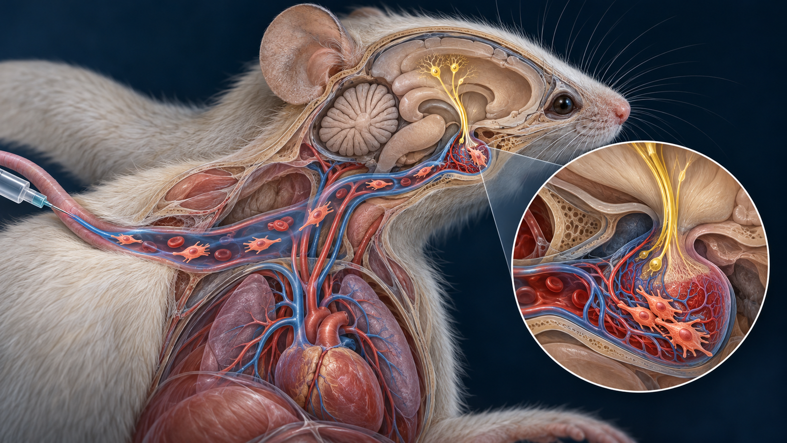
Generated editorial illustration of the study's proposed neurohypophyseal route. It does not depict measured biodistribution or brain imaging. Credit: CellXperience.
The transplanted cells in this study did not need to spread through the brain. They collected at a small vascular interface beneath it.
In a Science Advances paper published October 9, researchers gave human pluripotent stem cell-derived mesenchymal stromal cells intravenously to two autism-associated mouse models. Selected social, anxiety-related, and repetitive-behavior measures improved. The mechanistic trail led to oxytocin-producing neurons and the neurohypophysis, the posterior pituitary structure where those neurons release hormone into the circulation.
The paper links a peripheral cell infusion to a defined neural-endocrine circuit through several perturbation experiments. It remains a male-mouse study. Autism is heterogeneous, the assays are model-specific, and the work supplies no clinical evidence for treating autistic people.
Two infusions and a circuit-level question
The main experiments used male Shank3B knockout mice, a genetic model with altered social and repetitive behavior. Animals received one million hPSC-derived stromal cells by intravenous injection at three and seven weeks of age. Comparison groups received phosphate-buffered saline or human dermal fibroblasts, helping separate a cell-specific signal from the nonspecific effects of injecting human cells.
The stromal-cell group improved across selected three-chamber social, open-field anxiety, elevated-plus-maze, and grooming measures. Locomotor deficits did not recover. The result therefore depends on the endpoint rather than describing a global normalization of the animals.
Behavior alone could not identify where the cells acted. The authors tracked the infused cells and found their most notable accumulation around the neurohypophysis. This region sits outside the conventional blood-brain barrier and receives axon terminals from hypothalamic oxytocin neurons. It offers a plausible contact point between circulating stromal cells and a brain-linked endocrine system.
Markers of neuronal activation, including c-Fos and phosphorylated CREB, increased in oxytocin-producing regions. Oxytocin concentrations rose centrally and peripherally. These observations narrowed the proposed route: the infused cells appeared to act at a vascular neural-endocrine interface, followed by increased oxytocin signaling.
Perturbations make the route more credible
The team then interfered with the circuit at several positions. Chemogenetic inhibition of oxytocin neurons weakened the behavioral response to stromal cells. Activating those neurons produced overlapping effects. Blocking oxytocin receptors in the ventral tegmental area reduced parts of the response, placing a central reward and social-behavior circuit downstream of the endocrine signal.
No single perturbation proves the entire chain. Together, they make the proposed mechanism more specific than a familiar claim that MSCs exert broad anti-inflammatory or trophic effects. The paper identifies a physical destination, a neuronal population, a signaling molecule, and a downstream receptor site.
The cell-derived mediator was investigated through prostaglandin E2. Knocking down PTGS1 and PTGES, two genes required for PGE2 production, reduced the cells' capacity to activate the oxytocin pathway and weakened behavioral effects. The perturbation supports PGE2 as a contributor. Residual activity and the secretory complexity of stromal cells leave room for other mediators.
Direct electrophysiology or single-neuron measurements could strengthen the bridge between PGE2 exposure at the neurohypophysis and altered oxytocin-neuron firing. The reported evidence is convergent, though it does not isolate every cellular handoff.
A second model reveals heterogeneity
The researchers also tested BTBR mice, a separate inbred model used for autism-related behavioral research. The response varied. A subgroup with higher baseline oxytocin improved after stromal-cell treatment, while animals with lower oxytocin did not show the same pattern.
This split is intriguing because it points toward a biological condition for response. It is also post hoc. The threshold was derived after observing the data, and the subgroup needs prospective replication before it can function as a predictive biomarker.
The heterogeneity matters for the paper's central claim. If oxytocin tone influences whether the intervention changes behavior, averaging all treated animals could obscure the biology. A future study should define the threshold in advance, enroll enough animals to test it, and ask whether sex, age, stress, or model background alters the relationship.
The therapeutic distance remains large
Mouse social assays are experimental tools, not miniature versions of autistic life. Shank3B and BTBR mice capture limited biological and behavioral features. Neither represents the diversity of autistic people, co-occurring conditions, development, or support needs.
Only male mice were studied. Sex can affect oxytocin biology, immune signaling, and behavior, so that omission narrows the inference. The dosing also began early in postnatal development. Results could differ in mature animals, after one infusion, or with another stromal-cell product.
Systemic cell delivery brings safety questions that the behavioral experiments were not designed to settle. Biodistribution beyond the reported target, vascular trapping, inflammatory responses, product heterogeneity, persistence, and dose-dependent toxicity would need dedicated studies. hPSC-derived stromal cells add manufacturing questions about the starting line, differentiation consistency, residual pluripotent cells, genomic stability, and release testing.
The study's mechanistic precision should guide the next experiments. Replication across female and male animals comes first. A prospectively defined oxytocin-response analysis could test the proposed stratification. Pharmacokinetic work should establish when PGE2 changes, how long cells remain near the neurohypophysis, and whether a cell-free or shorter-lived intervention can reproduce the circuit effect.
The broader contribution is a route, not a clinical claim. A circulating stromal-cell product engaged a neural-endocrine interface in mice, and multiple perturbations tied selected behavioral effects to oxytocin signaling. The path from that result to any human intervention runs through replication, safety, respectful outcome selection, and clinical evidence that this paper does not provide.
Source and disclosures
This analysis uses the complete open Science Advances article, “HPSC-derived mesenchymal stromal cells ameliorate murine autistic-like phenotypes through activating oxytocinergic neurons”, published October 9, 2026. The study reports Chinese national, provincial, municipal, and scientific-talent funding. The authors declare no competing interests. The paper's diagnostic wording is reproduced only in the linked title; CellXperience uses identity-first or neutral language in its analysis. The hero is an original generated editorial illustration, not primary evidence.