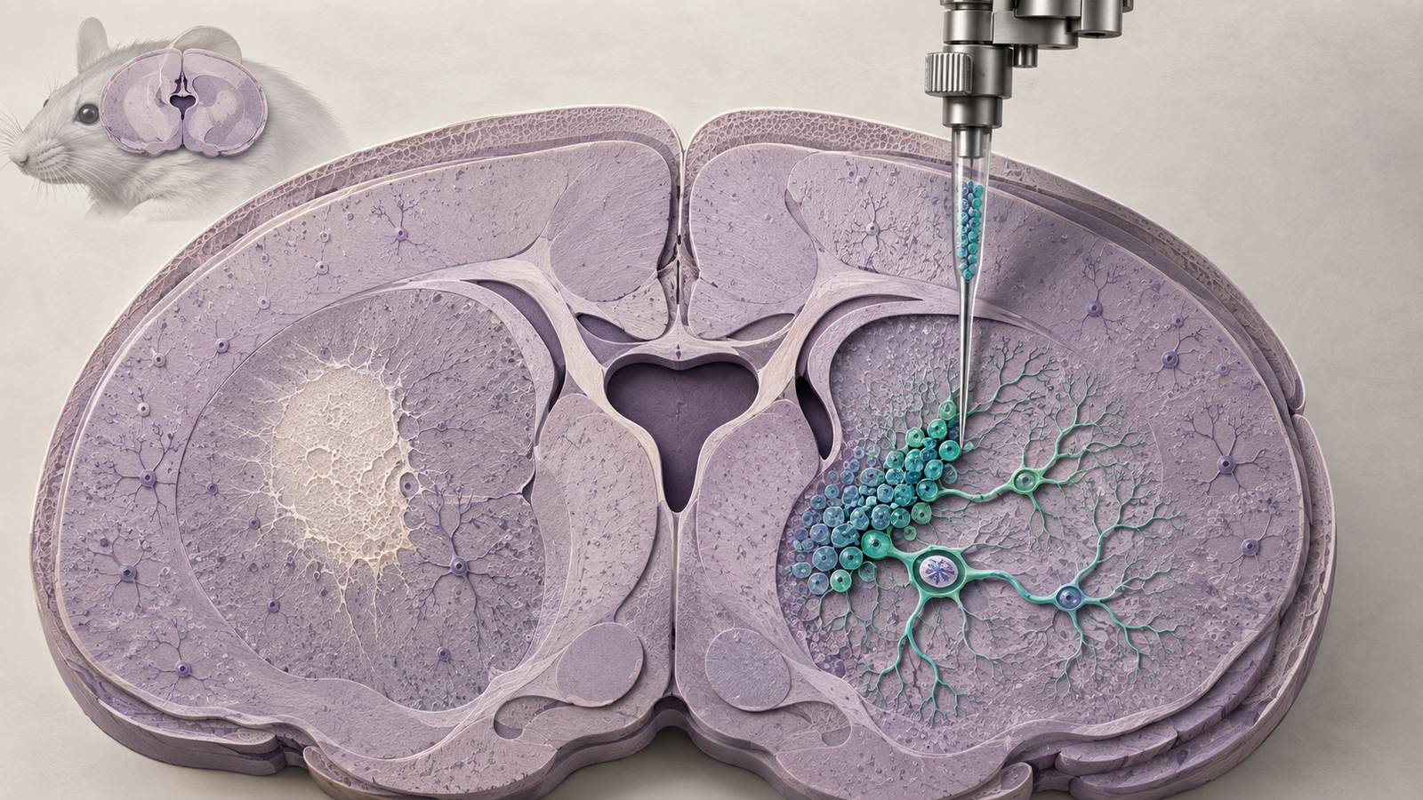
Editorial illustration of stereotactic neural stem-cell delivery beside an ischemic striatal lesion. Credit: CellXperience generated editorial illustration.
A neural stem-cell graft can survive and still miss its purpose. Cells may remain immature, adopt a glial fate, or influence damaged tissue through short-lived signals without becoming the neurons a replacement strategy was meant to supply. Counting grafted cells therefore answers only the first question.
An Advanced Science study attacks the fate problem directly. The researchers first mapped how neural stem cells change after ischemic stroke in mice. They identified Nrsn1 as a candidate regulator of neuronal commitment, connected it to the chromatin-remodeling protein Smarcc1, and then engineered a mouse neural stem-cell line before transplantation.
Four weeks after grafting, the Nrsn1-overexpressing cells produced more neuron-marker-positive and fewer astrocyte-marker-positive descendants than control-transduced cells. Motor tests and several tissue measures moved in the same direction. The experiment is genuinely useful because it compares two cellular products that differ in a defined fate-control mechanism. Its short follow-up and experimental cell line keep it far from a human stroke therapy.
Primary source
The complete peer-reviewed article was published online September 3, 2026. The full text, figures, methods, limitations, funding, and conflict statement were available for review.
The study begins with the injured striatum
The investigators used middle cerebral artery occlusion followed by reperfusion, a standard mouse model of ischemic stroke. Their analysis centered on the striatum, which sits near the subventricular zone where adult neural stem cells reside.
This choice creates a natural experiment. Injury can activate those endogenous stem cells. Some descendants move toward the damaged striatum, where their possible fates include neuronal and astrocytic lineages. The response is limited, but it gives researchers a place to search for the molecular decisions that separate one fate from another.
Single-nucleus RNA sequencing described gene expression at several recovery times. Single-nucleus ATAC sequencing measured accessible chromatin, and Stereo-seq preserved spatial information. Together, these methods placed quiescent and activated neural stem cells along inferred differentiation paths and located the relevant populations within the post-stroke tissue.
The maps showed an expansion of neuronal and astrocytic progenitor states around day seven. Nrsn1 was enriched along the path from neural stem cell to neuronal progenitor and appeared in DCX-positive cells in the striatum. That association supplied a candidate. It did not establish that Nrsn1 controlled the choice.
Increasing and decreasing Nrsn1 changes cell fate in culture
The next experiments moved into mouse neural stem-cell cultures. Oxygen-glucose deprivation followed by reoxygenation was used to mimic part of the ischemic environment. Nrsn1 expression rose as the cells increased neuronal-lineage markers.
The researchers then altered the gene in both directions. Stable Nrsn1 overexpression reduced the stem-cell marker Nestin and increased Pax6, Neurod1, Tubb3, and the proportion of Tuj1-positive cells. Two separate short-hairpin RNAs targeting Nrsn1 produced the opposite effect. Primary mouse neural stem cells supplied an additional culture system for parts of the observation.
Bidirectional perturbation makes the inference stronger than a sequencing correlation. The cultures still simplify the post-stroke niche. Oxygen-glucose deprivation does not reproduce blood flow, immune recruitment, extracellular matrix changes, or the mixture of dying and surviving cells in an injured brain.
The study also tested the endogenous compartment in vivo. An AAV2/9 vector under a Nestin promoter increased Nrsn1 in resident neural stem cells. Marker patterns shifted toward neuronal progenitors and mature neurons and away from astrocytes. Balance-beam, ladder-rung, and rotarod performance improved over four weeks, while MRI and Nissl staining favored the Nrsn1 group.
This AAV experiment and the later graft experiment ask related questions with different products. One changes resident cells already in the brain. The other changes cells before transplantation. Keeping those routes separate matters because their delivery risks, cell environments, and translational paths are not interchangeable.
Foxa2 and Smarcc1 give the fate shift a mechanism
Chromatin-accessibility data pointed to Foxa2 as an upstream transcription factor. A reporter containing the Nrsn1 promoter became more active when Foxa2 was expressed. Foxa2 also raised Nrsn1 RNA and protein, and the neuronal differentiation effect weakened when Nrsn1 was knocked down. That sequence places Nrsn1 downstream of Foxa2 in the culture system.
The more unusual connection came from an immunoprecipitation and mass-spectrometry screen. Smarcc1, also called BAF155, appeared among proteins associated with Nrsn1. Smarcc1 is part of the SWI/SNF chromatin-remodeling machinery that helps cells make lineage decisions by changing access to DNA.
Reciprocal pull-down experiments supported a physical association. Nrsn1 overexpression increased nuclear Smarcc1 without raising Smarcc1 messenger RNA. Knocking down Smarcc1 blocked the extra neuronal differentiation produced by Nrsn1. Fractionation and imaging placed more Smarcc1 in the nucleus when Nrsn1 was high and more in the cytoplasm when Nrsn1 was reduced.
Those results support a working chain: Foxa2 activates Nrsn1 transcription, Nrsn1 helps Smarcc1 accumulate in the nucleus, and Smarcc1-dependent chromatin regulation favors neuronal commitment. The paper did not identify the complete transport machinery or the relevant downstream chromatin targets. Protein-stability and proteasome experiments also failed to explain why total Smarcc1 protein increased. The mechanism has a useful middle, with both ends still open.
The graft comparison tests whether engineering survives contact with tissue
For transplantation, the team used C17.2 cells, an established mouse neural stem-cell line obtained from ATCC. A lentiviral construct produced stable Nrsn1 overexpression. Control cells carried a non-targeting sequence.
Each mouse received 500,000 cells by bilateral stereotactic injection into the subventricular zone, using a suspension of 100,000 cells per microliter. A separate PBS group was included in the broader procedure, while the central engineering comparison was Nrsn1-overexpressing C17.2 cells against control-transduced C17.2 cells.
At four weeks, MRI infarct-volume analysis used four animals per group, brain-weight analysis nine, behavioral tests ten, and dendritic and Nissl measures six. Histologic cell-fate quantification was reported from nine images per group. The paper presents data as mean plus or minus standard error and uses unpaired tests or one-way ANOVA as appropriate.
The engineered graft produced more GFP and NeuN double-positive cells and fewer GFP and GFAP double-positive cells. MAP2 staining, Nissl-positive cell counts, dendritic length, spine number, and projection complexity also favored the engineered group. On the balance beam, a difference emerged by two weeks. Ladder-rung performance separated at four weeks. Rotarod latency improved after transplantation and the engineered group maintained the stronger recovery.
The coherence across fate markers, tissue structure, and behavior is the paper's best feature. A single favorable stain would be easy to overread. Here, distinct measurements agree that Nrsn1 changed the graft and the host tissue response.
Better motor scores do not reveal which repair mechanism worked
The behavioral result cannot identify the route from cell engineering to recovery. Some GFP-positive graft descendants expressed neuronal markers, but marker expression does not prove mature electrophysiology, long-range axonal targeting, synaptic integration, or causal control of movement.
Nrsn1-overexpressing cells might also protect host neurons, alter inflammation, release trophic factors, or change the behavior of endogenous progenitors. The smaller infarct and healthier dendritic measures could reflect several of those processes at once. The study did not silence the grafted cells after recovery or use circuit tracing to show that their activity was necessary for the motor improvement.
Four weeks is particularly short for a replacement claim. A newly differentiating cell can express Tuj1, NeuN, or MAP2 well before its long-term identity and circuit function are settled. The same window cannot address late proliferation, unwanted migration, genetic stability, or durable graft behavior.
The paper's own limitations identify another important boundary. Young adult male mice were used for all treatment tests. Initial mapping included both sexes, but the therapeutic comparisons did not. Human stroke is concentrated in older adults, whose inflammatory, vascular, and regenerative environments differ substantially from a young mouse brain. Female and aged models are necessary, not decorative replication.
The cell product would have to change before clinical translation
C17.2 is an experimental mouse cell line, not a clinically manufactured human neural progenitor product. Stable lentiviral overexpression is useful for asking whether Nrsn1 can alter fate. A human program would need a defined donor source, manufacturing controls, potency assay, genomic and tumorigenicity testing, delivery strategy, and a decision about whether permanent Nrsn1 overexpression is desirable.
The target itself also needs calibration. Driving neuronal commitment too early can deplete a progenitor pool or reduce survival. Increasing a membrane-associated protein that influences chromatin regulation may affect cell behaviors beyond the measured lineage markers. The favorable dose of gene activity in C17.2 cells may not transfer to primary human neural progenitors.
The next persuasive experiment would use a well-characterized human iPSC- or ESC-derived neural progenitor, compare transient and stable Nrsn1 modulation, and follow aged animals for months. Lineage tracing should be paired with electrophysiology and circuit mapping. Selective inhibition of graft activity after behavioral recovery could then test whether the new cells are carrying information through the repaired circuit or helping the host brain by another route.