
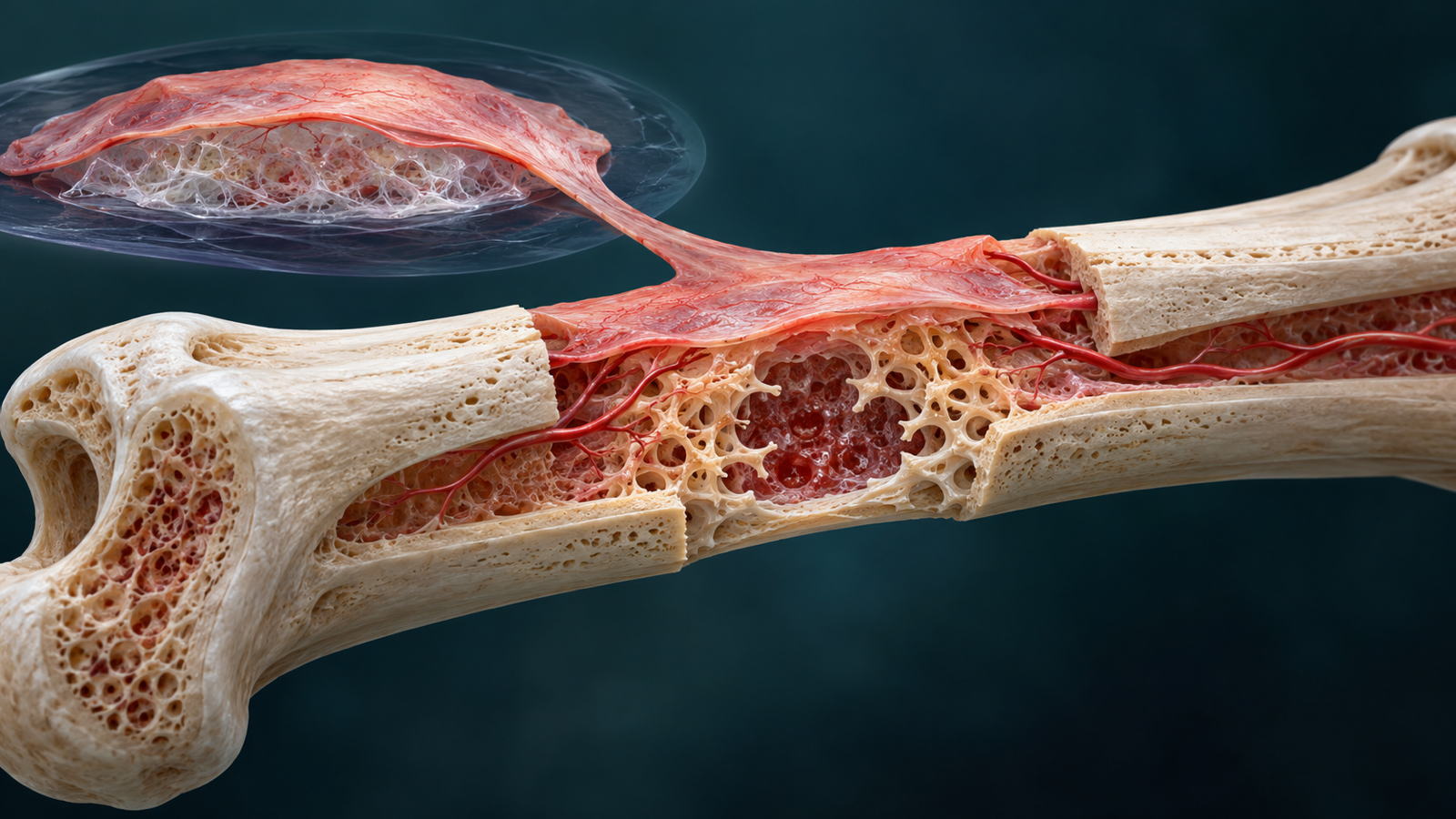
Generated editorial illustration of the culture-to-defect method. It is not study microscopy and does not depict a clinical procedure. Credit: CellXperience.
This organoid contracts before it grows bone.
In an open Advanced Science paper published October 22, researchers describe a long-lived culture made from the periosteum, the vascular membrane that wraps most bone surfaces. The culture can be replated through more than 20 passages and still form vascularized bone after transplantation. It also creates marrow-like spaces that host recipient cells.
Those results make the system interesting as a model of skeletal regeneration. A competitive transplantation experiment sets a firm boundary around the parallel claim that it supports hematopoietic stem cells. The organoid preserved several HSC-associated features during ex vivo co-culture, yet the recovered cells did not establish durable multilineage blood production.
The organoid is a layer around a shrinking dome
The starting material came from the femurs and tibias of six-week-old Sprague-Dawley rats. Investigators digested the periosteal tissue and used flow sorting to remove Desmin-positive muscle cells. The remaining population was embedded in Matrigel and grown in a mesenchymal expansion medium.
The structure differs from the compact epithelial spheres that often carry the organoid label. Periosteal cells moved toward the outside of the Matrigel dome and organized into a thin multilayered sheet. Cellular contraction and matrix remodeling reduced the dome’s average diameter from about six millimeters at plating to roughly three millimeters by day 14.
That geometry matters. The model recreates a tissue surface rather than a miniature bone. It also means part of the architecture comes from a large Matrigel scaffold whose composition and mechanical behavior are difficult to translate into a defined manufacturing process.
Cultures maintained their replating efficiency and growth kinetics through at least passage 20. Some assays extended to passage 30. Karyotyping at early and late passages and copy-number analysis did not reveal the abnormalities the authors tested for. These are encouraging stability checks, though they cannot exclude rare clones or every form of genomic change.
Long culture retains a mesenchymal core
Single-cell RNA sequencing covered 165,122 cells across fresh periosteum and organoids at multiple passages. Core mesenchymal and skeletal-lineage populations persisted. Immune and endothelial compartments present in the original tissue declined quickly in culture.
That shift is easy to miss if heterogeneity is treated as a single property. The organoids preserved several regenerative stromal populations while losing other elements of native periosteum. They are a durable mesenchymal reconstruction, not a complete replica of the source tissue.
Subcutaneous grafts in immunodeficient mice made the functional difference visible. The implants formed donor-derived cartilage and bone around cavities containing host blood and endothelial cells. Hematopoietic stem and progenitor phenotypes rose and fell during the eight-week experiment in a pattern consistent with formation of a marrow-like space.
The result shows that a stromal graft can organize host participation. It does not show that the organoid independently builds a complete marrow compartment, since the vascular and hematopoietic cells were recruited from the recipient.
The HSC test separates phenotype from function
The team also asked whether organoids could support hematopoietic stem cells outside the body. Mouse HSCs co-cultured with periosteal organoids retained surface-marker profiles, survival measures, lower reactive oxygen species, and lower senescence-associated signals better than several comparison conditions.
The decisive assay came afterward. Following 14 days of co-culture, 150 HSCs were mixed with competitor marrow and transplanted into lethally irradiated mice. All groups remained below five percent donor chimerism. The organoid-exposed cells produced no detectable multilineage reconstitution.
Surface phenotype and short-term fitness therefore did not translate into durable blood-forming function. This negative result is unusually useful. It warns against calling a niche functional solely because cultured cells keep an HSC-like immunophenotype or appear less stressed.
The experiment also identifies a concrete optimization target. A revised culture should be judged by serial blood-lineage output after transplantation, not by a larger collection of ex vivo markers.
Bone repair survives long passage
The skeletal results were stronger. In a four-millimeter critical-sized rat femoral defect, one organoid was placed into the gap. Early-passage and passage-25 grafts increased new bone and vascularization compared with Matrigel controls. A five-millimeter calvarial defect produced a similar pattern.
The study also compared intact organoids with dissociated cells at equal cell numbers. Both formed similar cartilage at an early subcutaneous time point, and freshly isolated periosteal cells could also make bone. The intact three-dimensional topology was therefore not required for every in vivo response.
That finding shifts the interpretation from a special organoid shape toward a preserved regenerative cell state. The culture may be valuable because it expands and maintains that state over time. Whether the final implant must remain intact will depend on the defect, delivery method, and desired host interaction.
The rat repair studies did not track donor cells, because graft and recipient were from the same species and lacked a lineage label. Histology can show new bone in the treated defect without assigning every osteogenic contribution to the transplanted cells. Host recruitment may account for part of the effect.
Zfp575 reveals a hidden three-dimensional state
Transcriptomic comparisons highlighted Zfp575, a poorly characterized zinc-finger transcription factor. Its expression was retained in the three-dimensional organoids and diminished in conventional two-dimensional culture.
Knocking down Zfp575 produced a useful mismatch between appearance and performance. The cultures still looked broadly intact in vitro, while their ability to make bone after transplantation fell. A standard morphology check would have missed the loss.
This makes Zfp575 a candidate marker of osteogenic competence rather than a validated release assay. The study did not prospectively set a threshold and test whether it predicts performance across new donors, batches, or human material. That is the work needed before the factor can guide manufacturing decisions.
What would move the platform toward translation
The current system starts with primary rat cells and relies on Matrigel. Repair experiments used small groups, short endpoints, and investigators who were not blinded. The study reports no human periosteal cultures, large-animal defects, load-bearing functional tests, biodistribution, or long-term tumor assessment. Sex-specific effects were not analyzed.
A translational version would need a defined matrix, human donor material, a reproducible cell-composition specification, and potency assays tied to durable repair. Mechanical strength and integration under normal loading matter more than histologic bone volume alone. Donor tracking would separate graft contribution from host recruitment.
For marrow support, competitive and serial transplantation should remain the gate. The current article earns attention because it presents attractive niche markers beside the assay that contradicts them. Bone repair is the credible lead. Durable HSC maintenance remains an unsolved part of the platform.
Source and disclosures
This analysis uses the complete open Advanced Science article, “Long-Term Expandable Periosteal Organoids Reconstruct a Functional Regenerative Niche That Support Hematopoietic Stem Cell Maintenance and Bone Repair”, published online October 22, 2026, including the full methods, results, and supplementary references available through PubMed Central. The authors report Chinese public and institutional funding and declare no conflicts of interest. The hero is an original generated editorial illustration, not primary evidence.