
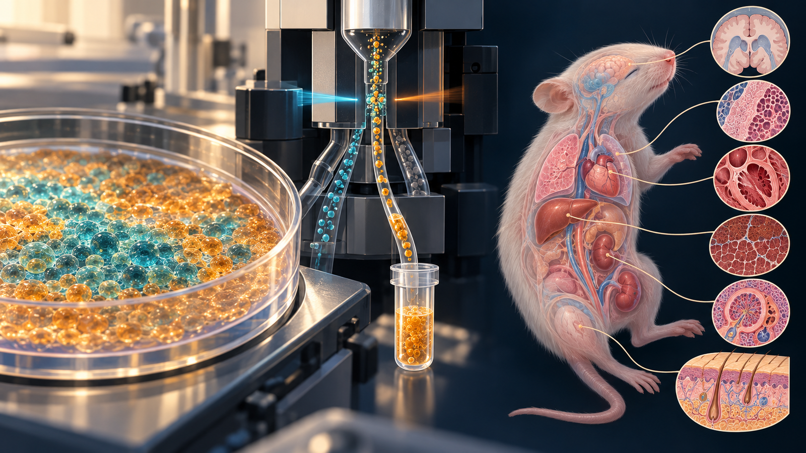
Generated editorial illustration of the study's progenitor-isolation and multi-organ engraftment method. It does not show measured microscopy or study animals. Credit: CellXperience.
The useful cell in a macrophage transplant may be the one that has not yet become a macrophage.
That possibility sits at the center of a Developmental Cell paper published online on October 6. Researchers found a yolk-sac-like myeloid progenitor inside a pluripotent stem cell differentiation culture, isolated a population enriched for that state, and tested its capacity to occupy empty tissue-resident macrophage niches. In specialized mice, descendants appeared in the brain, heart, kidney, liver, lung, and skin. Their gene-expression programs shifted with the organ in which they settled.
The experiment moves macrophage replacement closer to a defined starting population. It does not yet show disease correction. The recipients were neonatal, immunodeficient, or genetically depleted of resident macrophages, which made unusually permissive homes for human cells.
A transient state inside a mixed culture
Many tissue-resident macrophages begin during embryonic development, before definitive blood formation takes over. Yolk-sac progenitors seed organs early and then mature under local instruction. Microglia in the brain and Kupffer cells in the liver are familiar examples, though each tissue imposes a distinct identity.
The team used its MIGRATE differentiation protocol to generate human myeloid progenitors from pluripotent stem cells. Single-cell RNA sequencing and CITE-seq of 10,228 high-quality cells from six independent inductions mapped the developing culture. At day 18, an average 29.6 percent of cells occupied a transcriptional state that resembled human embryonic yolk-sac myeloid progenitors.
Those cells sat within a continuum. Their neighbors ranged from early progenitors to more mature macrophage-like cells, and no single marker drew a perfect border. The combination that proved most useful was high CD48 with absent CD163 inside the CD45-positive population. CD48 enriched the early state; CD163 tracked more mature macrophage features. Spectral flow cytometry across four PSC lines supported the pattern.
This is a practical result because mixed differentiations make cell identity hard to control. A transplant lot described only as “myeloid” can contain cells at very different developmental stages. Surface markers give the lab a way to enrich the state before transplantation and test whether developmental timing changes engraftment.
Earlier cells gain an advantage in the brain
For the brain experiment, the researchers injected cells intracranially into postnatal-day-4 immunodeficient mice carrying human CSF1. The host setting supports human myeloid survival. It also places the graft into a young brain whose resident niches and developmental signals differ from those of an adult patient.
Timing mattered. Day-18 cultures produced about 25 percent brain engraftment three to four weeks later, compared with 6.9 percent for day-25 cultures and 4.5 percent for day-32 cultures. The earlier window therefore contained something that the increasingly mature cultures were losing.
Sorting made the contrast sharper. Mice received 500,000 cells from either the total CD45-positive population, the CD48-high/CD163-negative progenitor-enriched fraction, or a CD48-low/CD163-positive macrophage-enriched fraction. Across seven experiments, the groups included 15, 18, and 21 animals, respectively. The progenitor-enriched fraction yielded roughly three times more human brain engraftment than the total CD45-positive population. The macrophage-enriched fraction performed worse.
The graft descendants did not remain generic myeloid cells. They adopted microglial and border-associated macrophage programs, and their states overlapped with profiles from primary human brain macrophages. That finding joins developmental identity to an in vivo fate rather than relying only on marker expression in a dish.
One delivery route, several organ identities
The wider test used FIRE mice, which lack tissue-resident macrophages in several organs because of a disruption in a regulatory element of the Csf1r gene. Newborn mice received about three million day-18 human myeloid progenitors by intraperitoneal injection. Three to five months later, human cells were detected in brain, heart, kidney, liver, lung, and skin.
Single-cell sequencing captured 2,083 human cells from brain, 347 from heart, 3,096 from kidney, and 201 from liver, plus a proliferating macrophage cluster. The dataset came from 20 tissues across six independently transplanted animals. Brain cells acquired microglial programs; liver cells included a MARCO- and TIMD4-positive Kupffer-like population; kidney and heart cells separated into their own tissue-associated states.
That organ adaptation is the paper's most compelling result. The same input culture did not produce one fixed macrophage phenotype everywhere. A developmentally early population responded to local tissue cues after arrival.
The counts also expose unevenness. Some tissues supplied thousands of recovered human cells, while others supplied hundreds. Detection and transcriptional resemblance do not establish that every graft-derived population performed the full functional repertoire of the native macrophage it resembled.
The niche did part of the work
Both mouse systems removed barriers that a clinical product would face. The brain hosts were immunodeficient and expressed human CSF1. FIRE mice offered vacancies because several endogenous macrophage compartments were depleted. Cells were delivered to newborns, when tissues are still developing and may be more accommodating than adult organs.
These choices are appropriate for asking whether the progenitors possess broad potential. They leave a different question unanswered: can the cells enter a mature organ whose macrophage compartment is already occupied, compete without damaging resident immunity, and improve a disease process?
Macrophage depletion itself could change tissue signals. Xenogeneic ligand-receptor mismatches could suppress some human programs while exaggerating others. The authors also caution that the CD48-high/CD163-negative gate enriches a state within a continuum. It does not produce a perfectly pure, indivisible progenitor class, and other cells in the culture may contribute to engraftment.
Replacement needs a function test
The platform opens several therapeutic directions. Genetically corrected progenitors might repopulate macrophage compartments affected by inherited disorders. Engineered cells could deliver a missing enzyme or replace dysfunctional microglia. Disease-specific iPSC lines could reveal how a mutation behaves as the same precursor adapts to different tissues.
Each direction requires a harder experiment than the one reported here. The next step is an adult disease model with a defined functional deficit, a realistic conditioning strategy, and a comparison against mature macrophages or other relevant cell products. Persistence should be linked to a tissue task: debris handling, metabolic support, pathogen response, synaptic maintenance, or correction of a measurable disease phenotype.
Safety has several layers. A proliferative progenitor can expand and populate a niche, which is desirable until growth escapes control. Manufacturing would need limits for residual pluripotent cells, unwanted hematopoietic states, genomic abnormalities, and batch variation. Long follow-up must examine ectopic engraftment and inflammatory behavior, especially if cells are engineered.
The study earns its place because it identifies a transplantable developmental window and follows that window across organs. Its claim stops before therapy. Yolk-sac-like progenitors can seed permissive macrophage niches and learn local identities in mice; whether they can repair a diseased adult tissue remains the experiment that matters next.
Source and disclosures
This analysis uses the complete open Developmental Cell article, “Inducible human yolk sac myeloid progenitors model myeloid ontogeny and enable tissue-resident macrophage replacement in vivo”, available online October 6, 2026. The study reports public and nonprofit funding from European, Belgian, UK, Spanish, and US organizations. The disclosure section lists consulting, advisory, founder, shareholding, and scientific-collaboration relationships for two senior authors and states that those engagements do not conflict with the work. The paper also states that ChatGPT was used for proofreading and that the authors reviewed the manuscript. The hero is an original generated editorial illustration, not primary evidence.