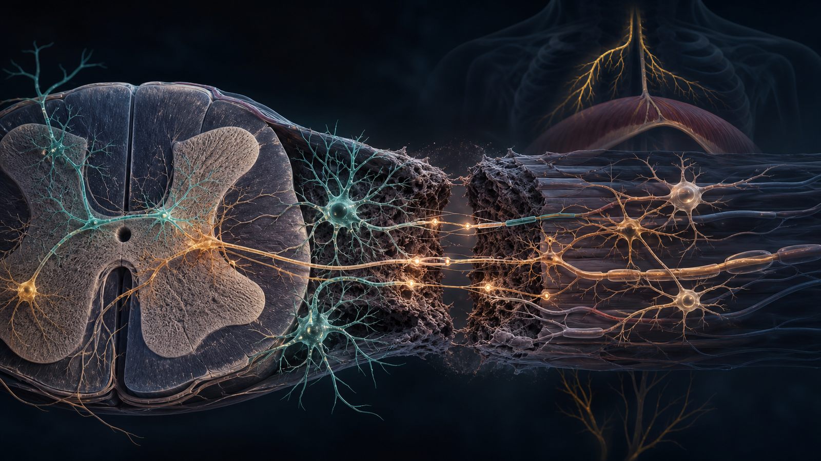
Editorial illustration of transplanted human spinal interneurons forming relay-like connections across an injured cervical spinal cord circuit. Credit: CellXperience generated editorial illustration.
Most stem-cell papers in spinal cord injury are easy to flatten into a familiar sentence: cells were transplanted, recovery improved in animals, more work is needed.
This new Science Translational Medicine paper deserves a more specific read. The interesting part is not simply that human cells survived in an injured rat spinal cord. The interesting part is the type of cell the authors chose, the circuit they used as the test case, and the way they tried to show host-to-graft and graft-to-host communication.
The study is still preclinical. It does not show that people with spinal cord injury can recover breathing or movement after this treatment. But it does make the repair question more concrete: can a defined human spinal interneuron population become part of a damaged motor circuit rather than merely sit near the injury and release helpful factors?
Primary sources
- Science Translational Medicine / PubMed: "Human spinal interneurons repair the injured rat spinal cord through synaptic integration" - https://pubmed.ncbi.nlm.nih.gov/42555754/
- Science Translational Medicine article page - https://www.science.org/doi/10.1126/scitranslmed.aea7461
- Gladstone Institutes summary - https://gladstone.org/news/engineered-human-neurons-rebuild-damaged-spinal-cord-circuits
Why V2a interneurons are a different bet
Spinal cord injury is not only a problem of missing tissue. It is a problem of broken circuitry. Motor, sensory, and autonomic pathways are interrupted, and the adult spinal cord does not naturally rebuild those lost connections in a clean, complete way.
The authors focused on V2a spinal interneurons. These are not dopamine neurons, retinal pigment epithelial cells, or a broad neural progenitor mixture. V2a interneurons are relay cells involved in motor control. In the context of cervical spinal cord injury, the authors used a breathing-related circuit as the proving ground because the phrenic motor network is anatomically and functionally tractable, and because high cervical injury can compromise diaphragm control.
That choice gives the paper its shape. Instead of asking only whether transplanted cells can survive after injury, the study asks whether engineered human spinal interneurons can connect into a circuit that has a measurable output.
The cells were generated from a human iPSC line engineered with channelrhodopsin-2, which allowed the researchers to use optogenetic stimulation as part of the functional testing. In culture, the team used multielectrode array recordings to show spontaneous and light-evoked activity and connectivity with spinal motor neurons.
Before transplantation, then, the cells could be made, matured, and shown to behave like electrically active neurons.
The transplant experiment was built around breathing
The in vivo experiment used a rat model of cervical spinal cord injury. The Gladstone summary describes transplantation into adult rats one week after injury, with assessment two months later. The institutional summary also notes that the team had worked toward cells that could be frozen and later thawed, a practical detail for any future translational version of the approach.
The paper's abstract describes several complementary readouts after transplantation. Pseudorabies-virus tracing from the diaphragm showed donor neurons synaptically connected with the injured phrenic motor network. Optogenetics and multiunit electrophysiology were then used to test functional communication in both directions: host-to-transplant and transplant-to-host.
The wording matters because "integration" can be a slippery word. In weaker usage, it can mean cells were still present and anatomically near host tissue. Here, the authors push for a stronger meaning: donor cells maturing into excitatory interneuron populations and taking part in a relay with injured motor networks.
The institutional summary gives the functional version of the same idea. When researchers activated the transplant site, diaphragm activity increased. When they activated the animals' own brainstem neurons, transplanted cells responded. Under ordinary conditions the breathing difference was subtle, but under low-oxygen or high-carbon-dioxide challenge, three-quarters of treated rats passed the stress challenge, while most untreated injured rats showed respiratory failure.
Here the study starts to feel alive scientifically. The cells are not presented only as a local biological dressing. They are being asked to help a circuit perform under stress.
What the paper can and cannot carry
The result is encouraging, but the translation path is still long.
A rat cervical injury model is not a human cervical injury clinic. The timing is early after injury, not months or years later. The target circuit is breathing-related, not the whole range of motor, sensory, autonomic, and hand-function priorities that people with cervical spinal cord injury live with. The cell population is defined and technically impressive, but it is not yet a product tested in people.
Those limitations keep the paper in the right category.
The paper's claim is narrower than "stem cells repair spinal cord injury": a defined human spinal interneuron population can be engineered from pluripotent cells, survive in an injured cervical spinal cord environment, mature into relevant neural and glial populations, connect with host circuitry, and support breathing-related motor output in a rat model.
That is a narrower claim. It is also a more interesting one.
Why this is more than a survival story
Survival is necessary for neural transplantation. It is only the first checkpoint. A cell that survives may still be irrelevant. It may fail to mature, fail to wire, wire incorrectly, or release factors without rebuilding the function that matters.
This study tries to move beyond that first checkpoint.
The authors report transplant maturation into multiple excitatory interneuron subtypes and glial populations. They also identify genes enriched in V2a spinal interneurons synapsing with the host phrenic motor network, including SLITRK5 and PLXNA3. That kind of molecular detail is useful because future development will need to know whether a graft contains the neuronal states most likely to connect productively.
The field has already learned, across several CNS cell-therapy areas, that "neural cells" is too broad a category. Product identity has to become sharper. A spinal interneuron program should eventually be judged by cell type, maturation state, purity, off-target populations, survival, circuit behavior, safety, manufacturability, and the disease setting in which the product is used.
This paper gives a more demanding preclinical template for that conversation.
The next questions are practical
Looking ahead, the Gladstone summary names two immediate translational questions. First, the approach needs to be tested in larger animals. Second, the team needs to learn whether it can work when treatment is delayed months or years after injury, not only shortly after trauma.
Those are not minor details. Many people living with spinal cord injury are not in the one-week post-injury window. A therapy that only works acutely would face a very different clinical path from one that can help chronic injury. The biology of the lesion, inflammation, scarring, spared circuitry, rehabilitation context, and surgical risk all change over time.
The circuit target also matters. Breathing is a powerful experimental system because it can be measured and challenged. But cervical spinal cord injury patients often prioritize arm and hand function, bladder and bowel function, pain, autonomic stability, and independence in daily activities. A repair strategy may need to prove itself one circuit at a time.
The paper works as a preclinical study because it shows a way to ask a better repair question.
How to read it now
This is a strong preclinical paper because it is specific.
The product concept is specific: human iPSC-derived V2a-enriched spinal interneurons. The disease model is specific: cervical spinal cord injury in rats. The circuit is specific: the phrenic motor network and breathing-related output. The mechanistic claim is specific: synaptic integration with injured host circuitry, supported by tracing, optogenetics, electrophysiology, and transcriptomics.
That specificity gives the field something more precise than hope and more actionable than a generic stem-cell transplantation result.
The next step is not to treat the paper like an early clinical promise. The next step is to see whether the same circuit logic survives tougher tests: larger animals, delayed treatment windows, scalable manufacturing, safety packages, and eventually a human trial design where the endpoint matches the circuit being repaired.
For now, the paper earns attention because it frames spinal cord repair as an engineering problem with a defined cellular part, a defined circuit, and a measurable output.